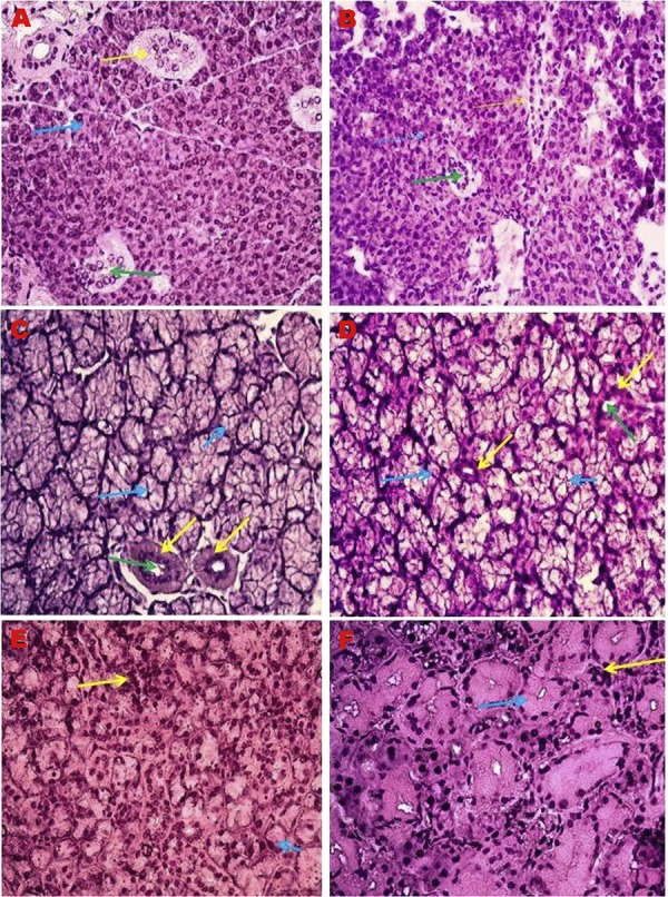
Figure 1. Major salivary gland histopathology H&E, 40X. A) Parotid gland of young mice. *Yellow arrow: Straited excretory duct; Blue arrow: Serous acini; Green arrow: Striated duct lumen; B) Parotid gland of old mice. * Yellow arrow: Straited excretory duct; Blue arrow: Serous acini; Green arrow: Striated duct lumen; C) Sublingual gland of young mice. * Yellow arrow: Straited excretory duct; Blue arrow: Mucous acini; Green arrow: Striated duct lumen; D) Sublingual gland of old mice. * Yellow arrow: Straited excretory duct; Blue arrow: Mucous acini; Green arrow: Striated duct lumen; E) Submandibular gland of young mice. *Yellow arrow: Serous acini; Blue arrow: Mucous acini; F) Submandibular gland of old mice; *Yellow arrow: Serous acini; Blue arrow: Mucous acini.

