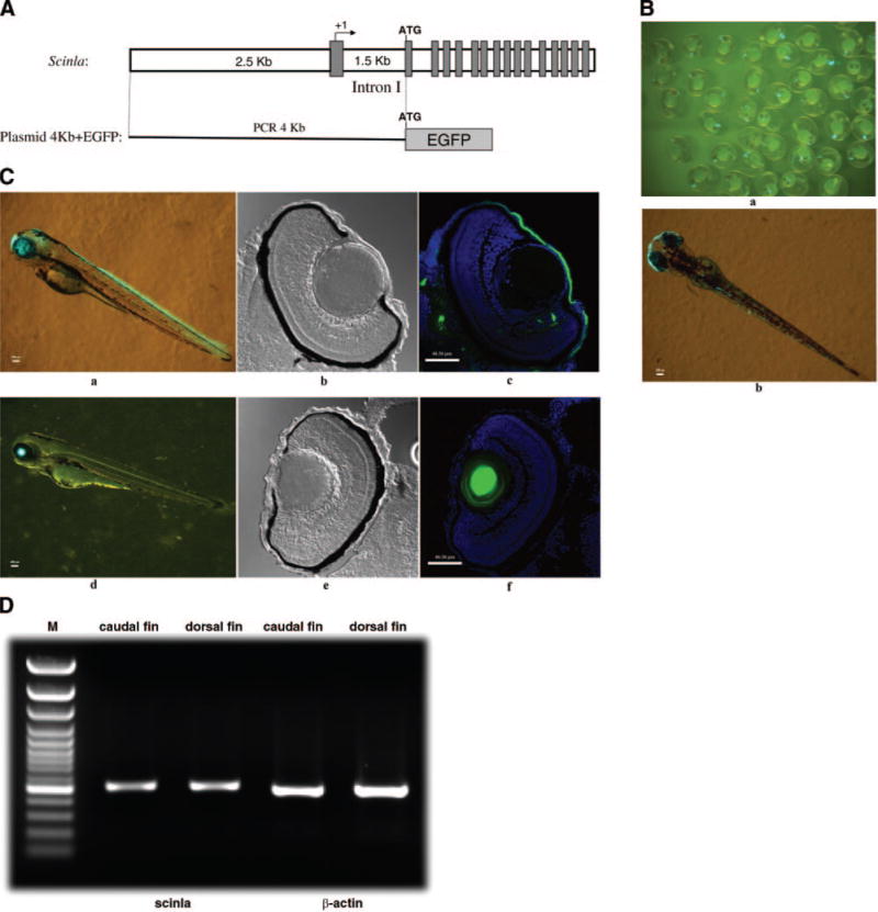
Figure 5.

A) Diagram of plasmid construct containing the zebrafish scinla 4 kb promoter fragment and the EGFP (4 kb + EGFP) fusion construct. The solid rectangles are exons. B) 1 dpf (a) and 3 dpf (b) zebrafish microinjected with the plasmid 4 kb + EGFP construct at the one-cell stage. C) 3 dpf stable transgenic zebrafish (a) and 3 dpf zebrafish microinjected with the αA-crystallin promoter driving the GFP transgene. Confocal DIC view of the 10 m-thick eye frozen section (b, e); merged view of DAPI staining and GFP view of 10 μm-thick eye frozen section (c, f). D) RT-PCR analyses of scinla expression in the dorsal and caudal fins using cDNA synthesized from RNA obtained from the specified tissues of adult zebrafish. β-Actin was used as a loading control.
