Figure 6.
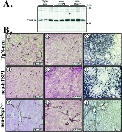
Evaluation of VEGF levels and distribution in tumors. (A) Equal protein amounts were analyzed by immunoblot with a specific antibody for VEGF. (B) Mammary tumor sections from TgN-neu (a–c), neu-hTSP1 (d–f), neu-tbsp1−/− (g–i) mice were evaluated for expression of VEGF by using two antibodies: GV39 M, which recognizes VEGF bound to VEGFR2 (b, e, h) and a polyclonal anti-mouse VEGF, which recognizes the receptor-free form (c, f, i). Staining with PECAM-specific antisera was used for identification of vessels (a, d, g).

