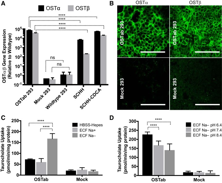
Fig. 2.
Expression and function of human OSTα/β in the established OSTab cell line. A: mRNA expression of human OSTα and OSTβ in Flp-In 293 cells transfected with OSTα/β vector (OSTab 293) compared with nontreated Flp-In 293 (wild-type 293) cells, Flp-in 293 cells transfected with empty vector (Mock 293), sandwich-cultured human hepatocytes (SCHH), and SCHH treated with a model bile acid, chenodeoxycholic acid (SCHH-CDCA, 100 μM, 24 h). Expression ratio of OSTα to OSTβ cannot be compared with the given normalization to wild-type. Values are means ± SD. Data were normalized to GAPDH and then compared with wild-type Flp-In 293 cells. B: immunofluorescence showing OSTα (green) and OSTβ (green) protein levels in OSTab and Mock cells. Scale bars = 100 µm. C and D: uptake of taurocholate (TCA) in OSTα/β-overexpressing Flp-In 293 cells (OSTab) and Flp-In 293 cells transfected with empty vector (Mock). C and D: Na+-free buffer and low-pH buffer increased OSTα/β-mediated uptake of TCA. OSTab and Mock cells were preincubated in HBSS-HEPES (pH 7.4) extracellular fluid (ECF) with sodium (ECF Na+, pH 7.4) or ECF in which sodium was substituted with choline (ECF Na−, pH 6.4, 7.4, and 8.4) at 37°C for 10 min. Uptake was initiated by addition of [3H]TCA or 3H-labeled estrone sulfate (300 nCi/ml, 20 µM, 2 min, 37°C) in the corresponding buffer. Values are means ± SD from 3 independent experiments. ****P = 0.0001; ns, not significant.

