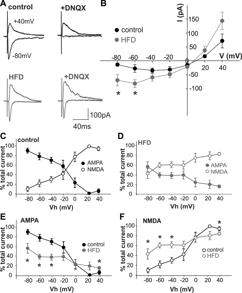
Fig. 4.
Acute high-fat diet (HFD) exposure alters the N-methyl-d-aspartate (NMDA): α-amino-3-hydroxy-5-methyl-4-isoxazolepropionic acid (AMPA) ratio in dorsal motor nucleus of the vagus (DMV) neurons. Glutamate currents were evoked electrically via stimulation of the adjacent nucleus tractus solitarius (NTS) with a bipolar electrode. Gastric-projecting DMV neurons were voltage clamped at potentials between −80 mV and +40 mV in the absence and presence of the non-NMDA receptor (R)-selective antagonist, 6,7-dinitroquinoxaline-2,3-dione (DNQX) (30 μM) to reveal the proportion of NMDA-R-mediated currents at each potential. A: representative evoked excitatory postsynaptic currents (eEPSCs) in control (top, black) and acute HFD (bottom, gray) gastric-projecting DMV neurons at −80 mV and +40 mV, in the absence (left) and presence (right) of DNQX. Note that, in control DMV neurons, DNQX almost abolished the eEPSC at −80 mV, suggesting that NMDA-R activation contributed little, if at all, to the glutamatergic eEPSC at this potential, whereas AMPA-R activation contributed little to the eEPSC at +40 mV. In contrast, the effects of DNQX to inhibit the eEPSC at −80 mV were attenuated significantly in acute HFD DMV neurons, suggesting that NMDA-R activation contributed significantly to the glutamatergic current at this potential. B: magnitude of the NMDA-R-mediated current in control and acute HFD DMV neurons was calculated at holding potentials between −80 mV and +40 mV to create a current-voltage relationship. Note that, in acute HFD DMV neurons, the NMDA-R-mediated current is increased significantly at potentials negative to −40 mV, suggesting a significant contribution to glutamatergic-mediated synaptic currents at rest (n = 3–8 neurons from 3 to 4 rats, respectively), *P < 0.05 vs. control (Student’s t-test). C: graphical representation of NMDA-R-mediated (○) and AMPA-R-mediated (●) currents as a percentage of total eEPSC at potentials between −80 mV and +40 mV in control gastric-projecting DMV neurons (n = 3–8 neurons, 3 rats). D: graphical representation of NMDA-R-mediated (○) and AMPA-R-mediated (●) currents as a percentage of total eEPSC at potentials between −80 mV and +40 mV in acute HFD gastric-projecting DMV neurons (n = 3–8 neurons, 4 rats). E: graphical representation of AMPA-R-mediated currents as a percentage of total eEPSC at potentials between −80 mV and +40 mV in control (black) and acute HFD (gray) gastric-projecting DMV neurons (n = 3–8 neurons for each, 3–4 rats). *P < 0.05 vs. control (Student’s t-test). F: graphical representation of NMDA-R-mediated currents as a percentage of total eEPSC at potentials between −80 mV and +40 mV in control (black) and acute HFD (gray) gastric-projecting DMV neurons (n = 3–8 neurons for each, 3–4 rats; *P < 0.05 vs. control (Student’s t-test).

