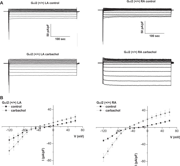
Fig. 1.
GIRK currents in atrial tissue. A: representative traces of currents measured in atrial myocytes isolated from the left and right atria of Gαi2+/+ mice. B: mean current-voltage relationships. Atrial myocytes were challenged with 10 μM carbachol. GIRK currents were larger in RA compared with LA (n = 10, 6 mice).

