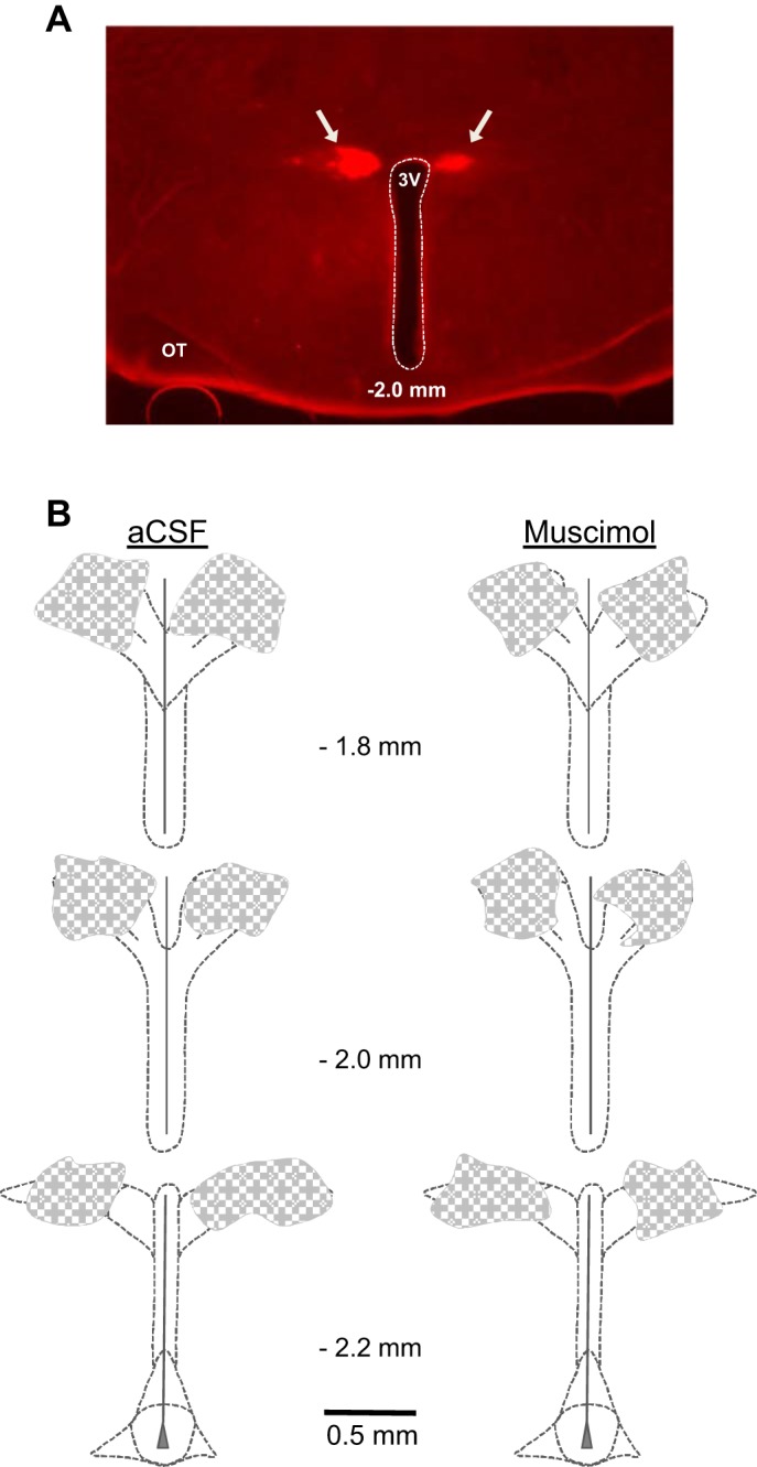
Fig. 5.

Location of PVN injection sites. A: histological section (50 µm thick) through the PVN at a rostral-mid plane showing representative distribution of rhodamine fluorescent nanospheres (arrows) marking sites of muscimol injection bilaterally into the PVN. B: summary representation of the location of aCSF (left; n = 5) and muscimol (right; n = 6) injections as indicated by the distribution of coinjected fluorescent nanospheres. Values in the center of each panel indicate the approximate rostral-caudal plane referenced to bregma. 3V, third cerebral ventricle; OT, optic tract.
