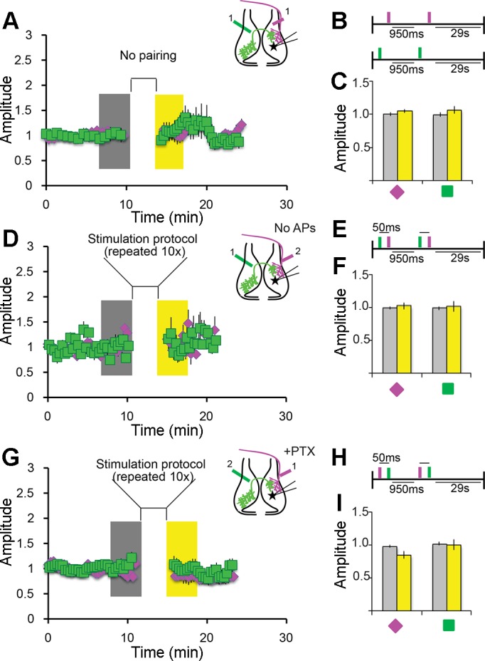
Fig. 4.

STDP depends on temporal proximity of input and postsynaptic depolarization. A–C: unpaired stimulation does not drive plasticity. A: bipolar electrode stimulation of IT (green squares) or RT (magenta diamonds) axons produced no plasticity. When stimuli were presented independently or with an interval >50 ms (data not shown), EPSC amplitudes were not significantly different from baseline. B: stimulation protocol presented as a schematic. C: summary data showing no potentiation. Data were collected as described in Fig. 3; n = 7 IT and 7 RT cells. D–F: postsynaptic depolarization is required for potentiation. D: a subset of cells recorded in the experiment represented in Fig. 3D failed to fire action potentials in response to RT or IT stimulation. No potentiation was observed in these cells. EPSCs were recorded and stimulation protocol was as described in Fig. 3, D–F. E: stimulation protocol. F: summary data showing no potentiation. Data were collected as in Fig. 3; n = 13 cells. G–I; inhibition is required for pairing-induced depression. G: RT stimulation before IT stimulation depressed IT responses (Fig. 3, G–I). PTX (100 µM) blocked the pairing-induced depression. H: pairing protocol as in Fig. 3H. I: summary data showing that the depression seen in Fig. 3, G–I is absent. Data were collected as in Fig. 3; n = 13 cells.
