Fig. 5.
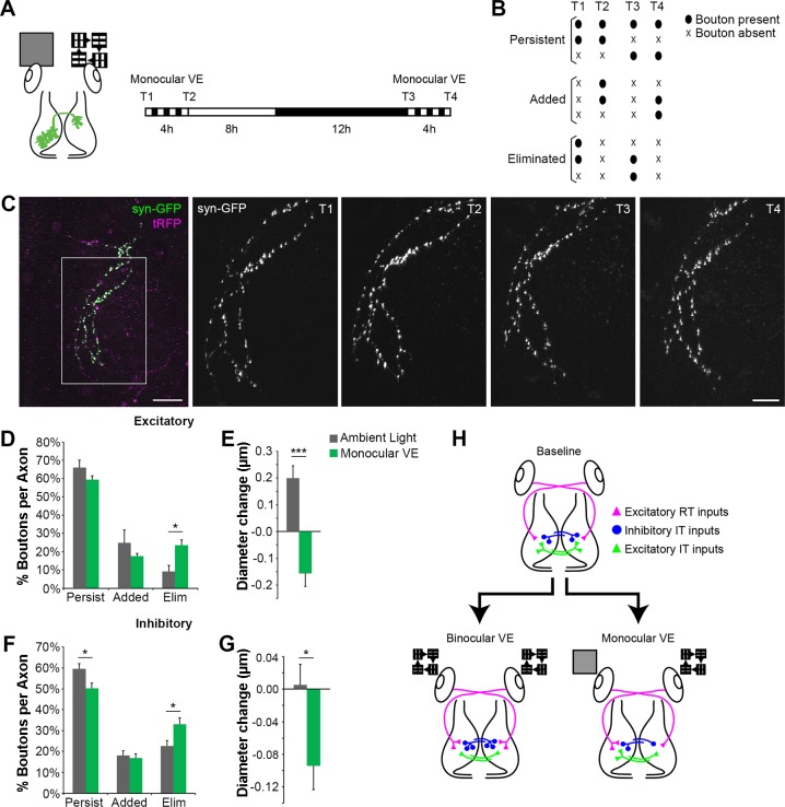
Monocular VE drives structural plasticity in intertectal axon boutons in vivo. A: experimental protocol. In vivo time-lapse 2-photon images of intertectal neuronal axons were collected before (T1) and after (T2) tadpoles were exposed to 4-h monocular VE provided to the eye contralateral to the neuronal cell bodies of the IT axons (schematized at left). Animals were returned to normal light-dark cycle. The next day, in vivo images were collected again before (T3) and after (T4) a second 4-h bout of monocular VE. This protocol tests the effect of visually driven activity on axonal outputs of IT-projecting tectal neurons. B: boutons were categorized as persistent, added, or eliminated in response to monocular VE as schematized. C: 2-photon Z projections of an IT axon expressing synaptophysin tagged with GFP (syn-GFP; green) and cytoplasmic turboRFP (tRFP; magenta). Scale bar: 50 µm. The boxed region was imaged repeatedly at higher magnification (right, T1–T4; scale bar: 25 µm). D and F: quantification of the percentage of intertectal boutons that persist, are added, or are eliminated during monocular VE. Monocular VE increases loss of excitatory (D) and inhibitory (F) boutons compared with ambient light. [excitatory (D): monocular VE, n = 6 axons; ambient light, n = 5 axons; *P = 0.014 eliminated, 2-tailed Student’s t-test; inhibitory (F): monocular VE, n = 11 axons; ambient light, n = 6 axons; *P = 0.040 persistent, *P = 0.038 eliminated, 2-tailed Student’s t-test]. E and G: quantification of the average change in diameter of persistent intertectal boutons. Persistent excitatory (E) and inhibitory (G) boutons shrink with monocular VE compared with ambient light [excitatory (E): monocular VE, n = 216 boutons; ambient light, n = 177 boutons; ***P < 0.0001, Mann-Whitney; inhibitory (G): monocular VE, n = 545 boutons; ambient light, n = 469 boutons; *P = 0.016, Mann-Whitney). Data are means ± SE. H: schematic illustrating the differences in structural plasticity of axon inputs after binocular VE and monocular VE. At baseline, each tectal hemisphere receives input from RT axons (magenta triangles), excitatory IT axons (green triangles), and inhibitory IT axons (blue circles). Binocular VE results in bilaterally symmetric addition of RT synapses and inhibitory IT synapses and elimination of excitatory IT synapses (bottom left). In contrast, monocular VE increases RT synapses in the tectum contralateral to the stimulus, and both excitatory and inhibitory IT neurons projecting axons from the stimulated tectum eliminate more synapses in the tectal hemisphere ipsilateral to the visual stimulus compared with ambient light. These data indicate that monocular VE produces bilaterally asymmetric structural plasticity. Data in binocular VE condition are from Gambrill et al. (2016).

