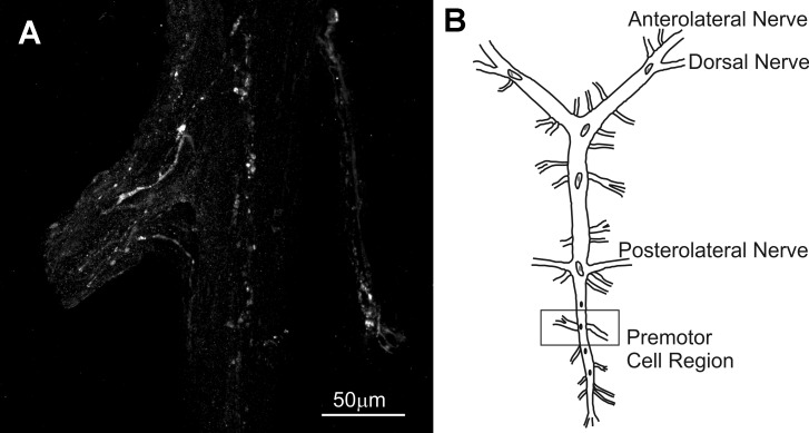
Fig. 1.
C-type allatostatin (AST-C) I/III-like immunoreactivity was present in the cardiac ganglion (CG). A: punctate nerve terminals, likely sites of peptide release, were located at several sites within the region of the CG, notably in regions in which the premotor neurons are located, suggesting that one or both of these peptides have access to these neurons. B: diagrammatic view of the CG; box indicates the region shown in A. This image is a maximum projection of 18 optical sections taken at 2-µm intervals.

