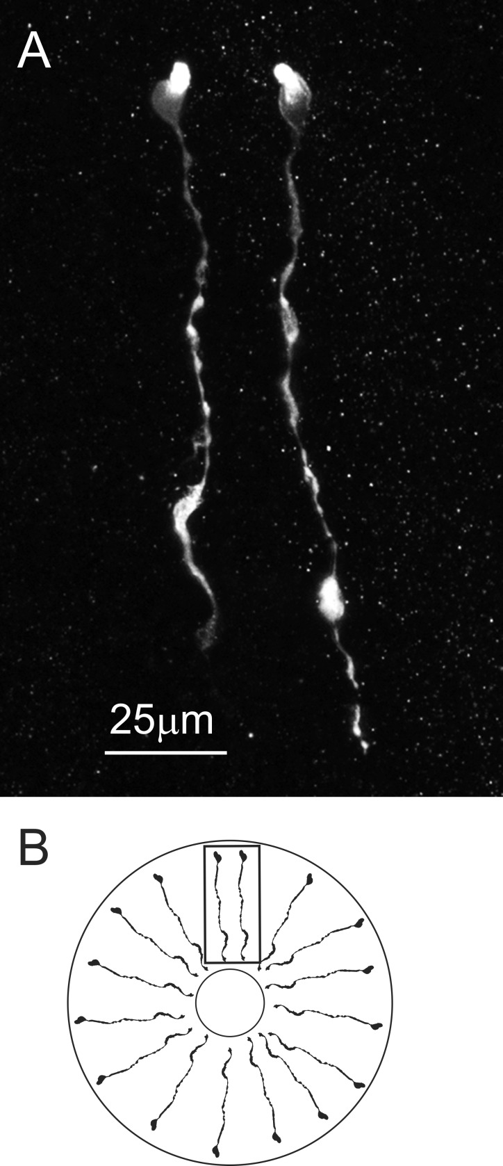
Fig. 3.
AST-C I/III-like immunoreactivity was present in epithelial endocrine cells in the posterior portion of the midgut. A: these immunopositive cells appear to span the epithelial layer and are morphologically similar to those previously described from the midgut of Cancer crabs (Christie et al. 2007). In the image shown, 2 AST-C I/III immunopositive cells are shown. This image is a maximum projection of 15 optical sections taken at 2-µm intervals. B: diagrammatic view of the midgut, showing the orientation of the AST-C I/III immunopositive midgut epithelial endocrine cells. Note that the drawing is not to scale: the lumen is shown much smaller than actual size. Box indicates orientation of image in A.

