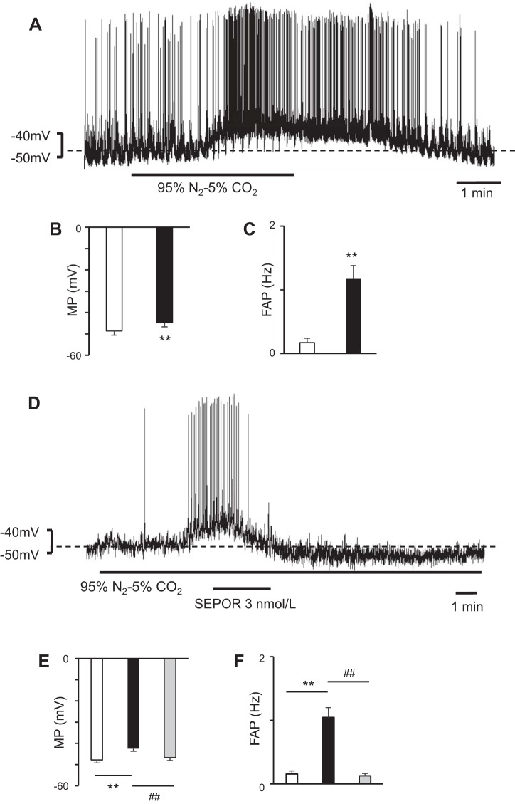
Fig. 3.
Hypoxia-depolarized bulbospinal rostral ventrolateral medulla (RVLM) neurons showing significant hyperpolarization during soluble erythropoietin (EPO) receptor (SEPOR) superfusion. A: neuron was depolarized during hypoxia. The hypoxia caused by bubbling 95% N2-5% CO2 depolarized the RVLM neuron. B and C: during hypoxia, the RVLM neurons were depolarized (B) and the frequency of action potential (FAP) in the RVLM neurons increased (C). Open bar, before hypoxia; solid bar, during hypoxia; values are means ± SE. **P < 0.01 vs. before hypoxia. D: addition of SEPOR hyperpolarized the hypoxia-depolarized RVLM neurons. In this case, the effect of SEPOR was very significant and prolonged. E and F: during SEPOR superfusion, the hypoxia-depolarized RVLM neurons were hyperpolarized (E) and the FAP in the RVLM neurons decreased (F). Open bar, before hypoxia; solid bar, during hypoxia; gray bar, during superfusion with SEPOR under hypoxic conditions; values are means ± SE. **P < 0.01 vs. before hypoxia. ##P < 0.01 vs. during hypoxia.

