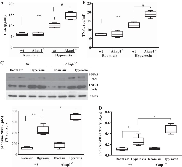
Fig. 4.
Akap1 genetic deletion increases inflammatory cytokine secretion into the BAL fluid, and increases phosphorylation and binding activity of NF-kB p65 in lung tissue after hyperoxia. Wild-type (wt) and Akap1−/− mice were exposed to room air or hyperoxia for 48 h, then BAL fluid was collected from each group and analyzed by ELISA for IL-6 (A) and TNFα (B) cytokine measurement. Data are shown as means ± SE (**P < 0.01 vs. wt room air; #P < 0.01 vs. wt hyperoxia; n = 5 mice/group). C: in separate experiments, total lung protein was collected and subjected to SDS-PAGE and Western blotting for phospho (P) and total (T) NF-kB p65 subunit. β-Actin served as loading control. Densitometry of phosphorylated NF-kB p65 shown below. Data are shown as means ± SE (**P < 0.01 vs. wt room air; *P < 0.05 vs. wt hyperoxia; n = 5 mice/group). D: analysis of DNA-binding activities of NF-kB p65 in lung nuclear extracts by TransAM NF-kB p65 activity assay kit. Data are shown as means ± SE (*P < 0.05 vs. wt room air; #P < 0.05 vs. wt hyperoxia; n = 5 mice/group).

