Fig. 9.
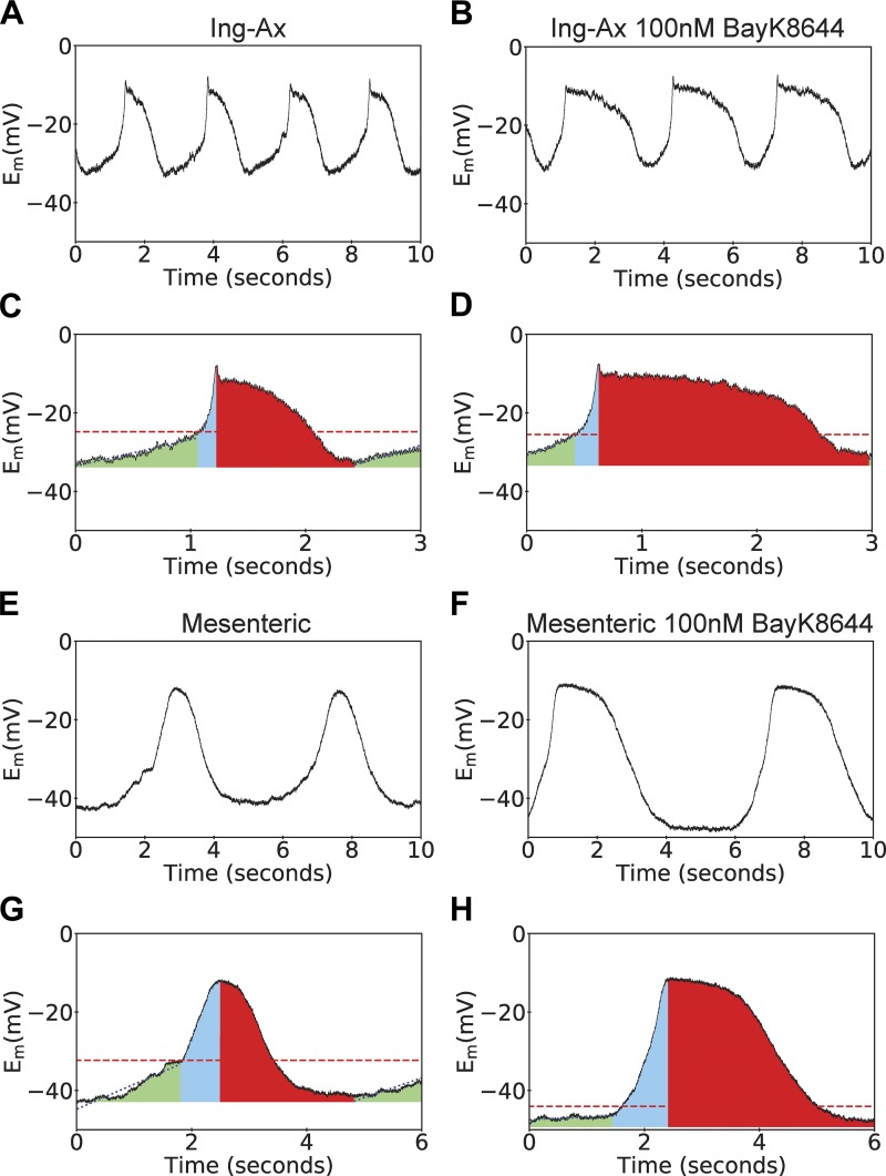
Membrane potential (Em) recordings of pressurized inguinal axillary (Ing-Ax) and mesenteric lymphatic vessels. Lymphatic contractions are driven by action potentials (APs) in isolated and pressurized murine lymphatic vessels. A and B: representative membrane potential traces over a 10-s period for one Ing-Ax vessel in Krebs buffer and after treatment with 100 nM Bay K8644 (BayK). C and D: colorized representation of Ing-Ax AP parameters analyzed on a per-AP basis in Krebs buffer and after treatment with 100 nM BayK. Linear diastolic depolarization period (green) reaches a threshold (dotted line), where the fast upstroke (blue area) reaches a peak, usually with a small spike of a few millivolts, followed by a plateau and repolarization phase (red area) to resting membrane potential. Mesenteric APs had a symmetrical, “hill-like” shape, as opposed to the classic AP observed in the Ing-Ax vessel. E–H: 10-s membrane potential traces from a single mesenteric vessel before and after BayK treatment, along with their individual AP analyses.

