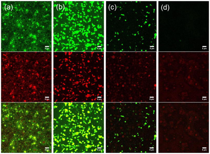
Figure 6.
Confocal microscopy images of calcein (green channel) and PNIPAM-RhB (red channel) co-loaded C-PS upon incubation in PBS with varying H2O2 concentrations at 37 °C for 24 h. From left to right: (a) PBS, (b) 163 mM (0.5%) H2O2, (c) 326 mM (1.0%) H2O2, (c) 652 mM (2.0%) H2O2. Scale bar = 5 µm.

