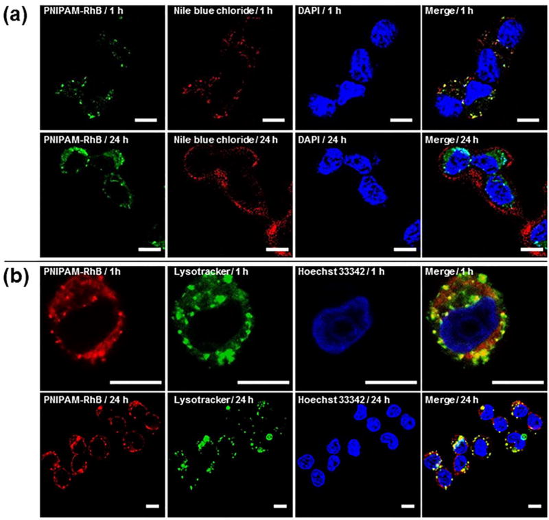
Figure 7.
Confocal fluorescence images of A2780 cells. (a) Cells were incubated with Nile blue chloride (red channel) and PNIPAM-Rh B (green channel) co-loaded C-PS for 1 h and 24 h. Nucleus were stained by DAPI. (b) Cells were first treated with PNIPAM-RhB loaded C-PS for 1 h, and then incubated with fresh medium for 1 h and 24 h. Nucleus and lysosome were stained by Hoechst 33342 and LysoTracker Green, respectively. Scale bar = 10 µm.

