Fig. 1.
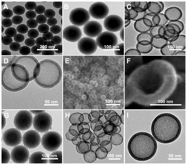
Electron microscopy images of the particles: TEM images of (A) Uniform c.a. 100 nm core Stöber NPs; (B) Disulfide-based mesoporous shell (15 nm) coated Stöber NPs synthesized by the addition of TEOS and BTESPD precursors; and (C and D) GSH-sensitive HMSiO2 NPs (c.a. 130 nm) under two magnifications. SEM images of (E) GSH-sensitive HMSiO2 NPs and (F) Broken NP showing voluminous (surface area: 446 m2 g−1) hollow interior. TEM images of (G) Mesoporous shell coated Stöber NPs synthesized by the addition of TEOS; (H and I) TEOS HMSiO2 NPs under two magnifications.

