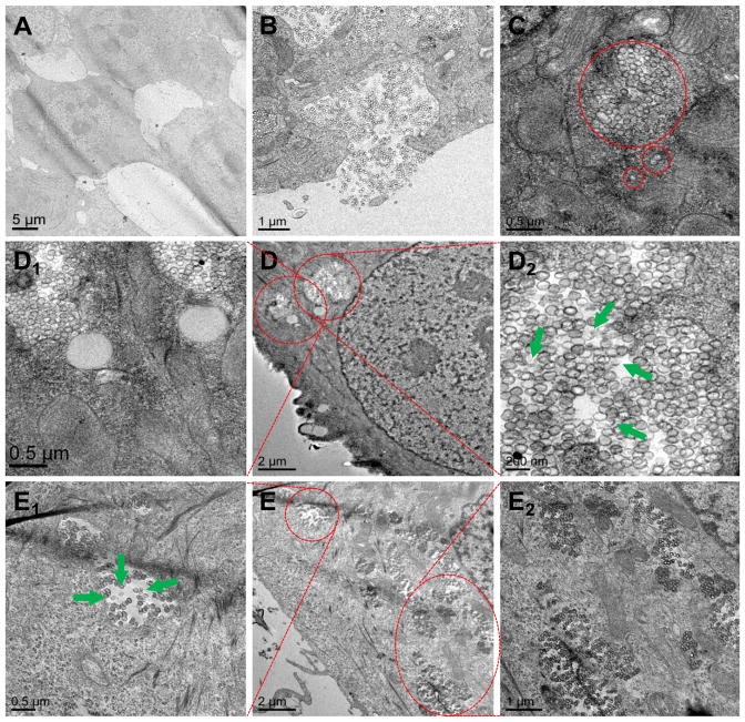
Fig. 6.
Representative TEM images for intracellular co-localization of GSH-sensitive HMSiO2 NPs in MCF-7 cells: (A) Control (cells without NP treatment); (B) Cells phagocytosing the hollow particles; (C) Particles are internalized in the endocytic compartments as an individual particle or as a group of particles; (D) Cells were treated with 50 μg mL−1 of NPs and incubated for 24 h; (D1 and D2) TEM images under higher magnifications. (E) Cells were treated with 250 μg mL−1 of NPs and incubated for 24 h; (E1 and E2) TEM images under higher magnifications. Green arrows show degraded particles in endolysosomal compartments.

