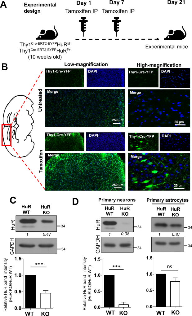
Fig. 1. Generation of neuron-specific HuR-deficient mice.
A. Tamoxifen was administered to conditional neuron–specific HuR-deficient (Thy1Cre-ERT2-EYFPHuRf/f) and age-matched control (Thy1Cre-ERT2-EYFPHuRf/+) mice at 10 and 11 weeks of age. Three weeks (21 days) after first tamoxifen injection, these mice (referred as experimental mice) were subjected to the experiments in Fig. 1B-C and Fig. 2 A–D. B. The representative confocal images of Cre-YFP in brain sections of neuron–specific HuR-deficient (Thy1Cre-ERT2-EYFPHuRf/f) mice either untreated or treated with tamoxifen as in A, Nuclei were stained with DAPI (blue). C. Western blot analysis of HuR in lysates from brain tissue of neuron–specific HuR-deficient (Thy1Cre-ERT2-EYFPHuRf/f) and control (Thy1Cre-ERT2-EYFPHuRf/+) mice after tamoxifen injection. Western blots were quantified by densitometry using ImageJ, error bars represent s.d. of biological replicates (N=15 mice for both KO and WT), ***p<0.05 by two-tailed Student’s t test. D. Western blot analysis of HuR in lysates from tamoxifen treated primary neurons and astrocytes isolated from HuR-deficient (Thy1Cre-ERT2-EYFPHuRf/f) and control (Thy1Cre-ERT2-EYFPHuRf/+) mice. Western blots were quantified by densitometry using ImageJ, error bars represent s.d. of biological replicates (N=15 mice for both KO and WT), ***p<0.001 by two-tailed Student’s t test.

