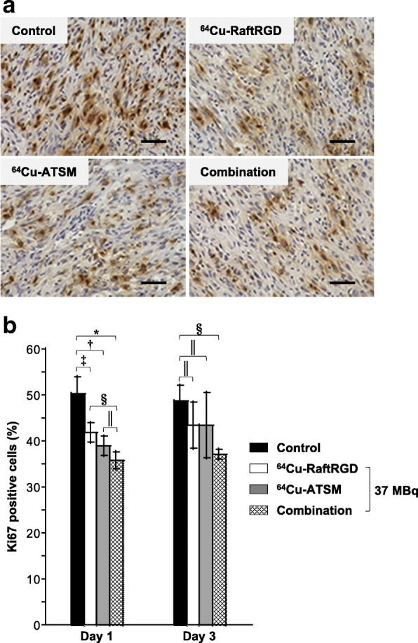
Fig. 4.

Ki67 staining (dark brown) of the tumor sections at days 1 and 3 p.i. of the vehicle solution (control) or 37 MBq of 64Cu-RaftRGD, 64Cu-ATSM, or a combination (18.5 MBq for each agent). a Representative images at day 1. Scale bar, 50 μm. Nuclei were lightly stained with hematoxylin (blue). b Quantitative analysis of Ki67 staining (the percentage of positively stained cells). n = 3−4/group; *, †, ‡, §P < 0.0001, 0.001, 0.01, and 0.05, respectively; ║P > 0.05
