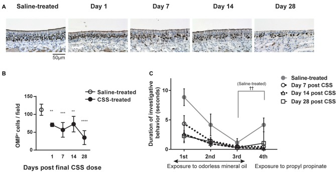
Figure 2.
(A) Immunohistochemical staining (brown) of olfactory marker protein-positive (OMP+) cells in the OE 1 day after the final dose of CSS. Mice received 20 doses (five cycles) of CSS. Data represent mean ± SEM (n = 6). **P < 0.01 (Mann-Whitney U test). (A) Representative images of immunohistochemical staining (brown) of OMP+ cells in the OE in saline-treated mice and on various days following the final CSS dose (n = 6). (B) Number of OMP+ olfactory receptor neurons (ORNs) per field in saline or CSS-treated mice. Data represent means ± SEM (n = 6). **P < 0.01; ***P < 0.001; ****P < 0.0001 compared with saline-treated mice (one-way ANOVA). (C) Olfactory habituation/dishabituation test. Mice were presented with a piece of filter paper soaked in odorless mineral oil three times for 3 min, at 1-min intervals. For the fourth exposure, the filter paper was soaked in the odorant propyl propionate instead of mineral oil. Data represent means ± SEM (n = 6). ††P < 0.01, third exposure compared with the fourth exposure (paired Student’s t-test). No significant differences were detected between the third and fourth exposures for CSS-treated mice on days 1–28, suggesting a decrease in olfactory sensitivity. CSS, cigarette smoke solution; OMP+, olfactory marker protein-positive.

