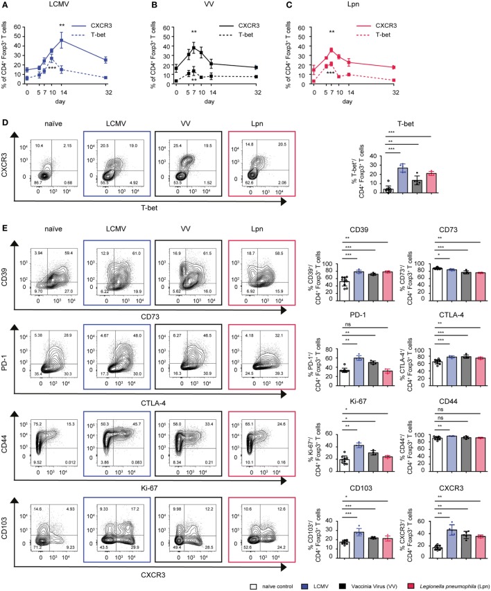
Figure 1.
CD4+Foxp3+ Treg cells specialize into T-bet+CXCR3+ Treg cells during Th1-dominated infections. C57BL/6 mice were acutely infected with LCMV WE (blue), Legionella pneumophila (Lpn, red), Vaccinia Virus (VV, black), or left naïve and expression of T-bet and CXCR3 (A–D) or the cell surface markers CD39, CD73, CTLA-4, PD-1, CD103, and activation/proliferation markers CD44 and Ki-67 (E) among live CD4+Foxp3+ Treg cells was determined by flow cytometry. (A–C) Frequencies of CD4+Foxp3+CXCR3+ or CD4+Foxp3+T-bet+ Treg cells over time and (D,E) peak expression levels of the indicated markers among splenic CD4+Foxp3+ Treg cells and representative FACS plots at the peak of activation (Lpn: days 5–7; VV: days 7–10; LCMV: days 10–14) or in naïve controls are depicted [mean ± SD, naïve: n = 10, Lpn, VV, LCMV: n = 4–5, biological replicates; plots display one representative of >3 independent experiments; *p < 0.05, **p < 0.01, ***p < 0.001 (Mann–Whitney test)].

