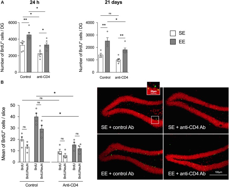
FIGURE 1.
CD4+ T cell depletion affects EE-induced neurogenesis in dentate gyrus of the hippocampus from SE and EE mice. (A) Chromogenic immunodetection of newborn BrdU+ cells measured in the DG 24 h after BrdU injection (left) or 21 days after the first BrdU injection (right) in mice raised in SE (white) or EE (gray). Mice were treated either with the depleting anti-CD4 or the control isotype antibodies (n = 4–6 mice per group). (B) Right Panel: confocal micrographs of the DG from control (left) or CD4+ T cell-depleted (right) mice housed in SE (top) or EE (bottom) showing the labeling of BrdU+ (green) and NeuN+ (red) cells. Left Panel: Histogram showing the mean number per slice (two slices labeled per hippocampus, n = 4 mice per group) of BrdU+ cells and BrdU+ NeuN+ cells.

