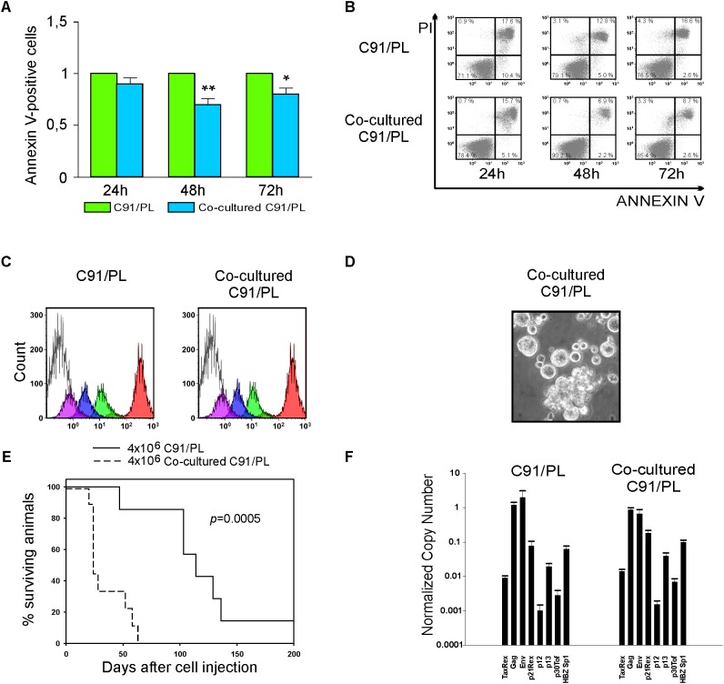
FIGURE 5.
Analysis of the in vitro crosstalk of C91/PL cells and HFF. (A) Co-culture with HFF reduced apoptosis in C91/PL cells. Apoptosis was analyzed in C91/PL cultured in standard conditions and in C91/PL cells co-cultured with HFF cells at 24, 48, and 72 h. Data are reported as ratio between the mean of the percentage of Annexin V–positive cells in two different co-cultures and the standard deviations (SD) of the ratio are calculated among four different experimental groups. Data are presented as mean ± SD. SD of the ratio was calculated according to the theory of error propagation. Statistical significance was determined by two-tailed Student’s t-test. ∗p < 0.05, ∗∗p < 0.01. (B) Flow cytometry analysis of Annexin V/Propidium Iodide (PI) stained C91/PL cells cultured in standard conditions (Upper) and in co-culture with HFF (Lower) at three different time points. A representative experiment is shown and the percentage of cells in each quadrant of the flow plots is provided. (C) Measurement of the in vitro proliferation of C91/PL cells cultured in the absence (Left) or presence (Right) of HFF. Cells were labeled with carboxyfluorescein succinimidyl ester (CFSE) and analyzed with XL Epic cytofluorimeter after 0 h (red), 24 h (green), 48 h (blue), and 72 h (violet). The shaded histogram shows the unlabeled cells. The plots show that co-culture with HFF does not affect the proliferation rate of C91/PL cells. Histograms represent the data obtained in two independent experiments. (D) Phase contrast images of C91/PL cells after co-culture with HFF. Eight-week-co-culture with HFF induced an increase in giant cells, thus resembling the C91/III cell line. Original magnification 100×. (E) Kaplan–Meier survival curves for 5-day-old NSG mice i.p. injected with 4 × 10⁶ C91/PL cells (seven mice) and with 4 × 10⁶ co-cultured C91/PL cells (nine mice). A statistically significant reduction (log-rank test; p = 0.0005) in the overall survival of mice injected with co-cultured cells was observed. (F) Analysis of HTLV-1 transcripts. Quantitative analyses of viral transcripts showed no significant variation in any of the HTLV-1 mRNAs in co-cultured C91/PL cells compared to the C91/PL cell line, suggesting that the increased survival and in vivo lymphoma induction acquired by co-cultured cells were not likely due to virus-encoded factors. Data are reported as described in Figure 3B.

