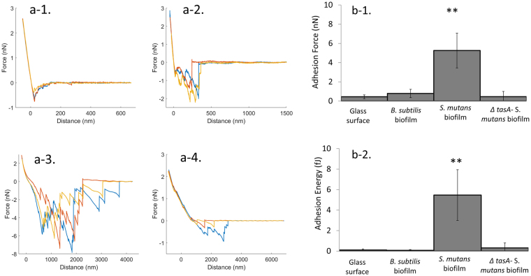
Figure 2.
B. subtilis cell interacts with S. mutans biofilm via TasA protein. B. subtilis bacterial cells were incubated on coverslip for 20 minutes until attachment to the glass. One B. subtilis cell was picked and attached to tipless cantilever coated with PDA. Single cell force spectroscopy measurements were taken using AFM. (a) representative force profiles of B. subtilis WT on a glass surface (a-1), B. subtilis WT on a B. subtilis biofilm (a-2), B. subtilis WT on S. mutans biofilm (a-3) and B. subtilis ∆tasA on S. muatns biofilm (a-4). (b) Adhesion force (b-1) and energy (b-2) of single B. subtilis cell to glass, B. subtilis biofilm and S. mutans biofilm. The adhesion force and energy between B. subtilis cell and S. mutans biofilm was significantly higher compare to glass. However, ∆tasA strain did not show a high adhesion force or energy towards S. mutans biofilm. The data are displayed as a mean value ± standard deviation. **P value < 0.01 compared to control.

