Figure 3.
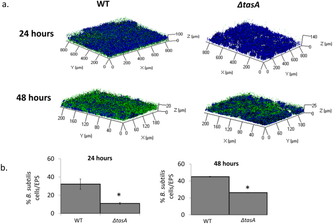
B. subtilis’ TasA protein effects the structure of co-species biofilm and the incorporation of B. subtilis into co-species biofilm. (a) Representing CLSM picture of co-species biofilm. B. subtilis (WT (YC161 marked with GFP) or ΔtasA (marked with GFP) and S. mutans were grown together for 24 h (upper panel) or 48 h (lower panel). S. mutans dextran-associated EP were marked in blue using Alexa fluor 647. In both of the time periods, there are more WT B. subtilis cells among the S. mutans’ dextran-associated EP compare to the ΔtasA strain. Moreover, after 48 h there is an increase in the amount of incorporated B. subtilis cells within the dual-species biofilm in both strains (WT and ΔtasA). (b) Quantification of the fluorescent intensity of GFP for live bacteria and Alexa fluor for EP. The data are displayed as a mean value of data from five biological repeats each performed as triplicate. The amount of WT B. subtilis cells is significantly higher than the ΔtasA strain after 24 and 48 h. Furthermore, there is an increase in the amount of B. subtilis cells within the dual-species biofilm in both of the strains. The data are displayed as a mean value ± standard deviation. *P value < 0.05 compared to WT.

