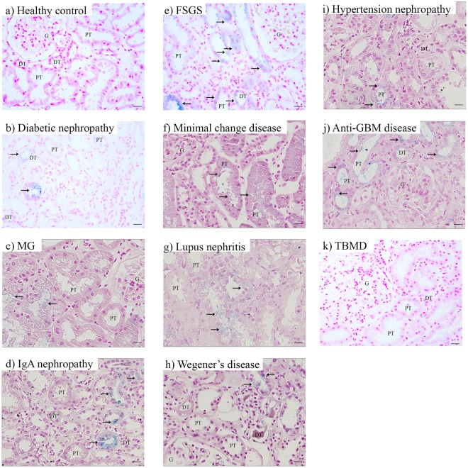
Figure 1.
Iron deposition in chronic kidney disease. Representative images of Perls’ staining in healthy control (a), diabetic nephropathy (b), membranous glomerulopathy (MG; c), IgA nephropathy (d), focal segmental glomerulosclerosis (FSGS; e), minimal change disease (f), lupus nephritis (g), Wegener’s disease (h), hypertension nephropathy (i), anti-glomerular basement membrane (GBM) disease (j), and thin basement membrane disease (TBMD; k). Renal structures indicated as glomerulus (G), proximal tubule (PT), distal tubule (DT). Iron indicated with arrows. Scale bar 20 µM.

