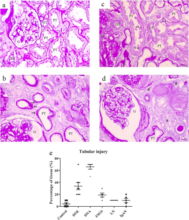
Figure 7.
PAS staining for tubular injury in chronic kidney disease. Representative images of PAS staining in control (a) or chronic kidney disease showing atrophic tubules with loss of brush border and enlarged basement membrane (b), blebbed tubules (c) and interstitial fibrosis (d). Percentage of tissue with tubular injury scored (E) per patient. Renal structures indicated as glomerulus (G), proximal tubule (PT), and distal tubule (DT). Symbols indicate proximal tubular brush border with asterisk, blebbed tubules with double asterisk, basement membrane with arrow, and fibrosis with hashtag. Scale bar 20 µM.

