Figure 5.
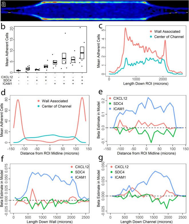
Spatial statistics of leukocyte adhesion as a function of coatings. (a) Leukocyte adhesion distribution heatmap for all ICAM-1 containing conditions in the straight channel device (Supplemental Fig. 1A). Flow direction is left to right. These consist of a length 4 expansion and an extended straight section to capture relaxation of the expansion effect. Wall associated adhesion peaks immediately after the initial expansion, and again at the collision-inducing point where the expansions end. Channel center adhesion subtly increases over the length of the channel. (b) Mean number of adherent cells per ROI in response to factorial combinations of CXCL12, SDC4, and ICAM1 in the coating, demonstrating additive effects. Each point represents mean numbers for a single participant, N = 6 donors. Boxes indicate mean and standard error. (c) Sub-ROI quantification using a mask for wall-associated vs. center of channel leukocytes as a function of flow distance along the ROI, demonstrating relaxation of the expansion effect for wall-associated but not channel center leukocytes. Wall associated adhesion probability peaks immediately after the expansion, then declines across the ROI until the junction with the constriction induces further collisions between erythrocytes and leukocytes, generating a second peak of adhesion. In contrast, channel center adhesion gradually increases across the length of the channel. (d) Validation of the sub-ROI quantification method by quantifying across the cross section of the ROI: Wall associated adhesion is restricted to the sidewalls, whereas channel center adhesion peaks in the channel center. (e) Linear model fits across the ROI cross section for the relative contributions of CXCL12, SDC4, and ICAM1 to adhesion. As expected, the contribution of adhesion molecule ICAM1 is most significant in the channel center, where shear stress is highest. (f) Linear model fits along the ROI for coating contributions to leukocyte adhesion on the sidewall. The initial spike of adhesion at the expansion is strongly ICAM-1 dependent. (g) Linear model fits along the ROI for leukocyte adhesion in the center of the channel. Adhesion immediately after the expansion is dependent on all three coatings, whereas ICAM-1 becomes more dominant as the expansion effect relaxes.

