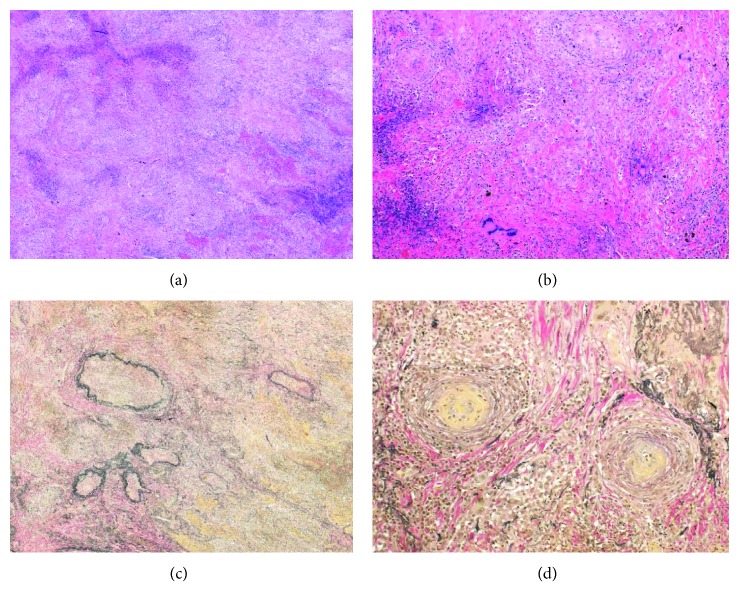
Figure 2.
Histopathological findings of lung nodules from our patient with granulomatosis with polyangiitis. (a) Hematoxylin and eosin (H&E) staining (×40) of lung biopsy showing necrosis and inflammatory cell infiltration. (b) H&E staining (×100) of lung biopsy showing vascular occlusion and multinucleated giant cells. (c) Elastica van Gieson (EVG) staining (×40) of the lung showing destruction of the arterial medium. (d) EVG staining (×100) of lung biopsy showing vascular occlusion.

