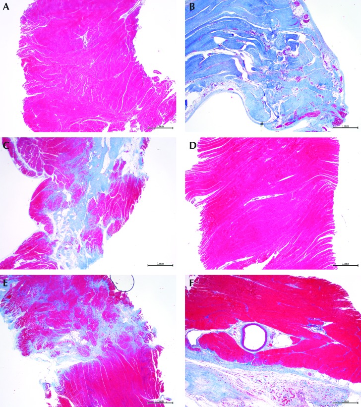
Figure 4.
Panels A through C represent the severe-infarction group; panels D through F represent animals with mild infarcts. (A and D) Grossly unaffected areas of the myocardium away from the infarct show no replacement fibrosis. (B) The infarcted area from a severely affected heart has extensive and transmural replacement fibrosis, whereas (E) the mildly affected heart demonstrates multifocal distribution in the infarcted area. (C) The border zone from the severely affected heart shows multifocal and poorly demarcated replacement fibrosis, whereas that from (D) the mildly affected group has less replacement of muscle. Masson trichrome stain (red, muscle; blue, collagen); magnification, 20×.

