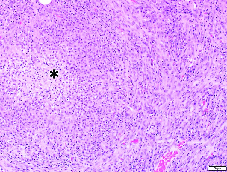
Figure 1.
Photomicrograph of peripancreatic adipose tissue. A core of necrotic debris and degenerate neutrophils (asterisk) is surrounded by plump, activated macrophages and a thin capsule of fibrous connective tissue mixed with neutrophils. Small amounts of granulation tissue are present. Hematoxylin and eosin stain.

