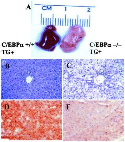
Figure 6.
(A–E) Histologic analysis of TG+, α−/−, and, TG+, α+/+ wild-type liver at age 7 days. (A) Gross appearance of TG+, α+/+ (Left) and TG+, α−/− (Right) liver at age 7 days. Note the yellow color of the TG+, α−/− liver (Right). (B and C) Hematoxylin/eosin stain of TG+, α+/+ (B) and TG+, α−/− (C) liver. The large vacuoles in the cytoplasm of the TG+, α−/− liver indicate fat storage. (D and E) Oil red O stain of TG+ wild-type (D), and TG+, α−/− liver (E). Note the large amount of lipid droplets in the TG+, α−/− liver.

