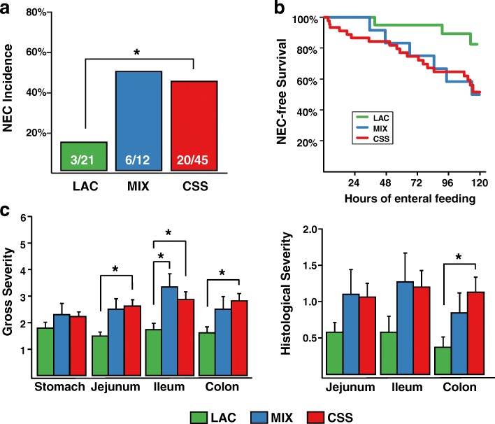
Fig. 1.
Phenotypic outcomes of the study. a NEC incidence within each of the three formula groups; Fisher’s exact test. b Kaplan-Meir analysis comparing survival time before euthanizing for NEC across the three formula groups; log-rank test. c Gross and histological NEC severity scores across GI regions for the three formula groups. For the gross severity, each of the GI regions is assessed at the time of dissection and assigned a score of 1–2 (healthy tissue), 3–4 (moderate inflammation), or 5–6 (pneumatosis and necrosis). For histological severity, H & E-stained tissue sections are scored as 0 (no damage), or from a range of 1 to 4 based on extent of necrosis, villus blunting, and pneumatosis. Values are mean +/− standard error of the mean; linear model with birthweight and farm as covariates and Tukey’s post hoc comparisons

