
Figure 2.
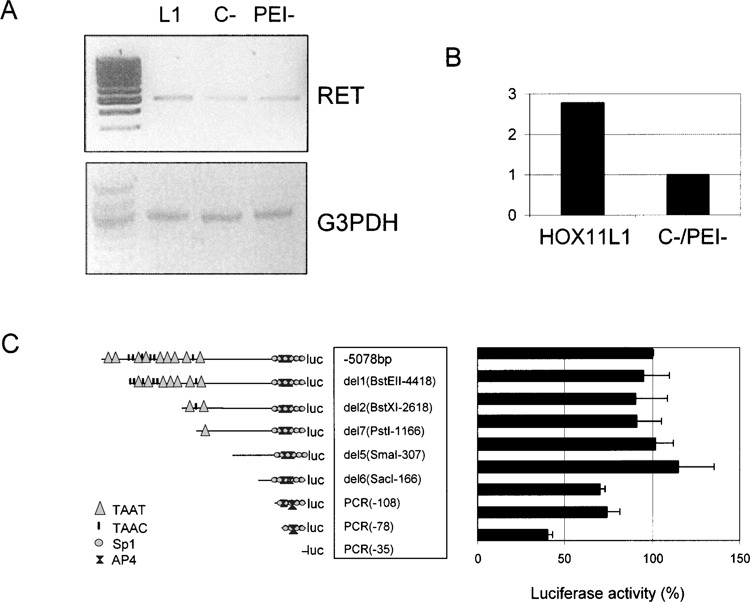
RET expression following transfection with HOX11L1. (A) SK-N-MC cells were transfected with pcDNA3.1TOPO-HOX11L1 (L1), with the empty vector (C−), and not transfected (PEI−). In any case, cells were treated with NaB to induce RET expression. RT-PCR was performed using a couple of primers designed on RET exon 18 and RET exon 20, respectively, and the amount of RET product compared to that obtained under the same condition for the GA3PDH gene. (B) Bands thus obtained were quantified to show the amount of RET expression in the presence and in the absence of HOX11L1. (C) Sequentially deleted reporter plasmids of the RET promoter, represented on the left, are reported along with corresponding HOX11L1 activations, plotted on the right as percentage of the activity of the full-length (−5078 bp) construct. Data represent the means ± SE (error bars) of at least three independent experiments performed in duplicate.