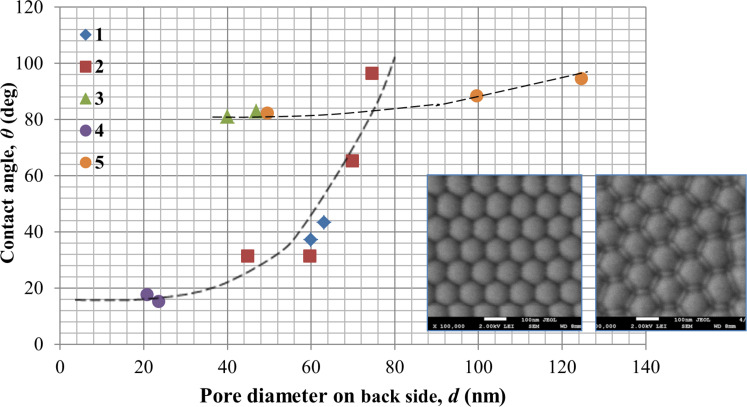
Figure 12.
Contact angle measurements for the back side of the PAM as a function of pore diameter for various membrane thicknesses: 1 (blue diamonds) – 28 µm; 2 (red squares) – 45 µm; 3 (green triangles) – 65 µm; 4 (purple circles) – 75 µm; 5 – contact angle as a function of cell diameter for the as-made PAM (before pore opening). The inset shows SEM images of the back surface before (left) and after (right) chemical etching. The black dotted lines are polynomial fits shown as a guide to the eye for selected data sets.

