Figure 7.
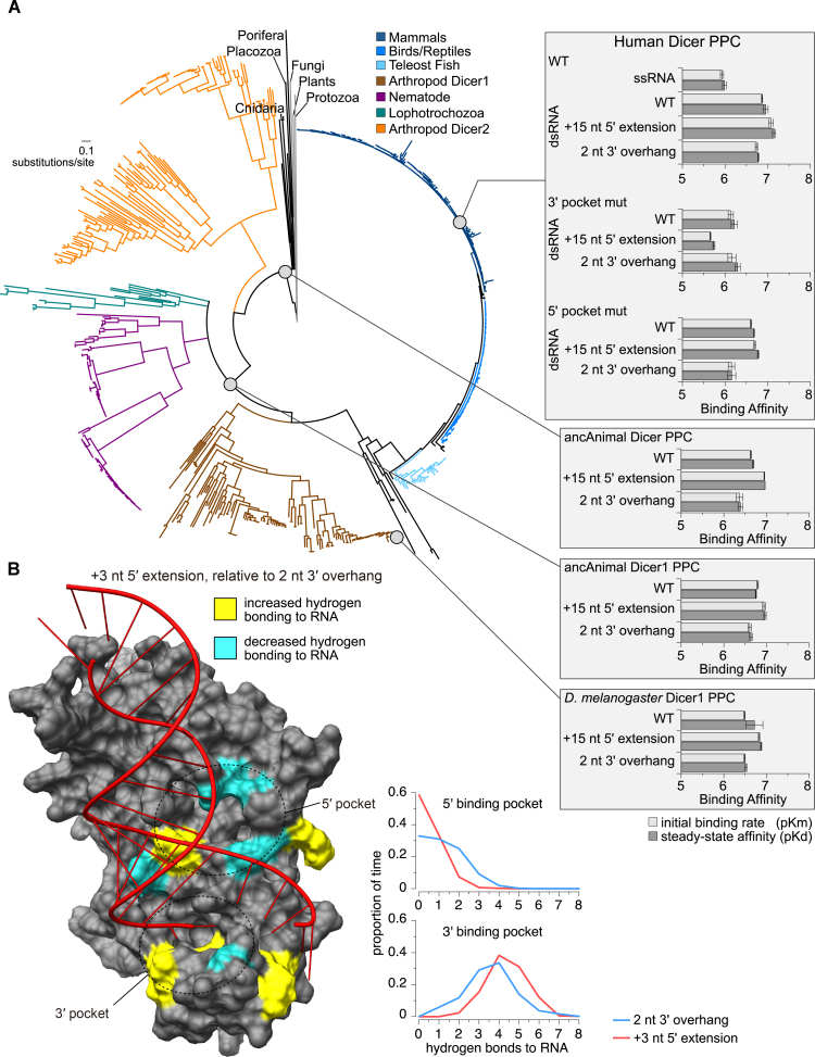
Animal Dicer Platform-PAZ-Connector (PPC) domain shows conserved affinity for dsRNAs with 5′ extension. (A) Reconstructed maximum-likelihood consensus phylogeny of the animal Dicer protein family (shown at left, with major taxonomic groups color-coded). Affinities of ancestral and extant Dicer PPC domain (gray circles) for dsRNAs of miR-HSUR4 were measured using a label-free kinetics assay (shown in right insets). The plots show log-transformed initial binding rates (pKm; light gray) and steady-state dissociation constants (pKd; dark gray), with standard errors indicated. (B) Replicate molecular dynamics simulations of Human Dicer PPC (PDB ID 4NHA) bound to dsRNA having a 2 nt 3′ overhang or a +3 nt 5′ extension. We calculated the proportion of time samples for which each amino acid residue formed hydrogen bonds with each RNA ligand. Only the +3 nt 5′-extended dsRNA is shown in the structure. Residues showing significantly increased (yellow) or decreased (cyan) hydrogen bonding to the 5′-extended dsRNA are highlighted in the structure (P< 0.001). Graphs show the proportion of time samples over 10 ns simulations for which each number of hydrogen bonds were observed between the Dicer PPC 5′ pocket (top) or 3′ pocket (bottom) and its dsRNA ligand, averaged over three independent simulations.

