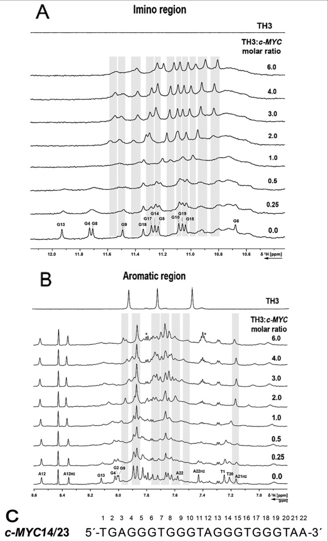
Figure 4.
(A) Imino and (B) aromatic region of 1D 1H NMR spectrum of the c-MYC14/23 G-quadruplex DNA with increasing [TH3]:[DNA] ratio and ligand alone. The spectra were recorded at 298 K, 700 MHz. Experimental conditions: 100 μM DNA in 25 mM Tris•HCl (pH 7.4) buffer containing 100 mM KCl in 10% d6-DMSO/90% H2O. (C) Sequence of c-MYC14/23 with the numbering used for the partial assignment of DNA signals. Signals marked with a star are arising from ligand self-aggregation (∼10%, Supplementary Figure S16) after transferring in buffer during the titration. These aggregated species do not interact with the DNA (Supplementary Figure S17).

