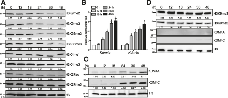
Figure 1.
Up-regulation of KDM4A and KDM4C and reduction of H3K9me2/me3 is found following stimulation of B cells by Tfh-cell derived signals. (A) Levels of histone markers detected by immunoblotting (IB) with nuclear extracts from [HEL + anti-CD40 + IL-21]-stimulated splenic B cells from MD4 mice at indicated time points. (B, C) Kdm4a and Kdm4c mRNA (B) and protein (C) levels at indicated time points in stimulated splenic B cells isolated from MD4 mice. Lamin B was used as the protein loading control in (C). (D) Levels of H3K9me2, H3K9me3, KDM4A, and KDM4C detected by IB with nuclear extracts from LPS (2.5 μg/ml) stimulated splenic B cells from C57BL/6 mice at indicated time points. H3 is served as the loading control. The relative levels of indicated proteins in (A) (C) and (D) after quantification were indicated. Results in (B) represent the mean ± SEM (n = 3). **P < 0.01, ***P < 0.005 (Student's t-test).

