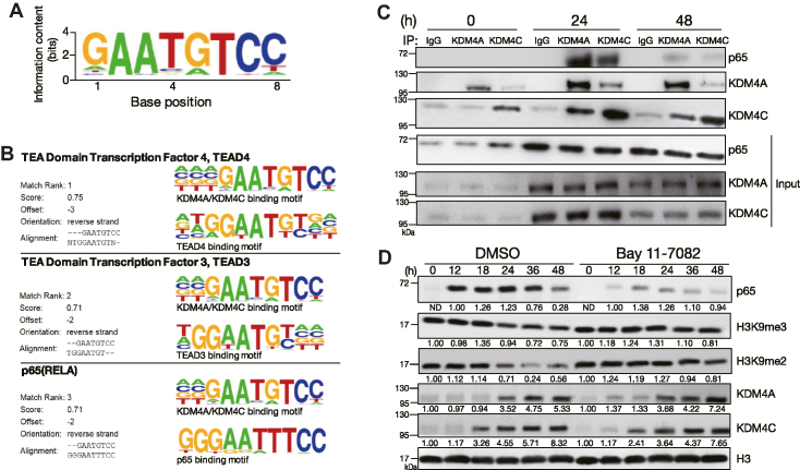
Figure 3.
KDM4A, KDM4C and NF-κB temporarily form a complex during B cell activation. (A) Consensus KDM4A and KDM4C-binding motif identified using the de novo motif-discovery program Homer Software, P = 1 × 10−42. (B) Analysis of de novo discovery of ChIP-seq peaks derived from the common motif of KDM4A and KDM4C binding sites revealed the top three transcription factor binding motifs. (C) Co-immunoprecipitation (co-IP) using nuclear extracts from MD4 mouse splenic B cells stimulated with HEL + anti-CD40 + IL-21, followed by immunoblot showing the interaction of NF-κB p65 with KDM4A and KDM4C 24 h after stimulation. Rabbit IgG was used as the control antibody in the immunoprecipitation. (D) Immunoblot and quantitative analysis showing the levels of indicated nuclear proteins in stimulated MD4 mouse splenic B cells treated with 15 μM of BAY 11-7082 NF-κB inhibitor at indicated time points. Relative levels of indicated proteins in (D) after quantification were indicated.

