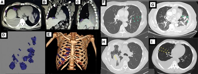
Figure 1.
Volumetric assessment in a patient with right-sided mesothelioma. A–C) Segmentation of the tumor in axial, coronal, and sagittal planes is shown. D–E) Tumor volume and relative distribution in the thorax are shown. Axial computed tomography images of patients with malignant pleural mesothelioma showing (F) no fissural involvement, (G) fissural involvement, (H) maximal fissural thickness (Fmax) of 56 mm, and (I) Fmax of 5 mm.

