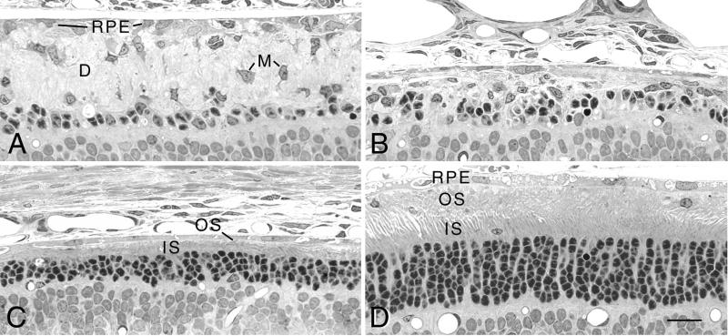
Figure 2.
Light micrographs of a single RCS rat retina injected in the superior hemisphere with Ad-Mertk at P22 and taken at P52. (A) Peripheral retina in the inferior hemisphere shows a thick OS debris zone (D) containing numerous macrophages (M). (B) Posterior retina in the inferior hemisphere. (C) Posterior retina in the superior hemisphere. (D) Peripheral retina in the superior hemisphere in the region of Ad-Mertk injection. (Bar = 20 μm.)

