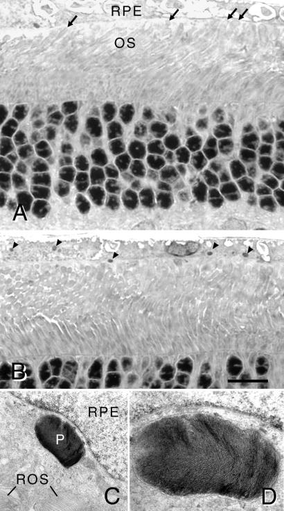
Figure 4.
(A and B) Light micrographs of retinal sections of an RCS rat at P52 that received stock Ad-Mertk at P22. (A) Tips of rod OS (ROS) reach the apical surface of the RPE (arrows). (B) Numerous phagosomes are present in the RPE cytoplasm (arrowheads), demonstrating ingestion of shed ROS membranes. (Bar = 15 μm.) (C and D) Electron micrographs of a retinal section comparable to that in B. (C) A large phagosome (P) is found in the cytoplasm and near the nucleus of an RPE cell. Tips of ROS are apposed to the apical surface of the RPE cell. ×10,625. (D) Higher magnification (×28,600) of a serial section to (C) illustrating ROS-like lamellar disk membranes within the phagosome.

