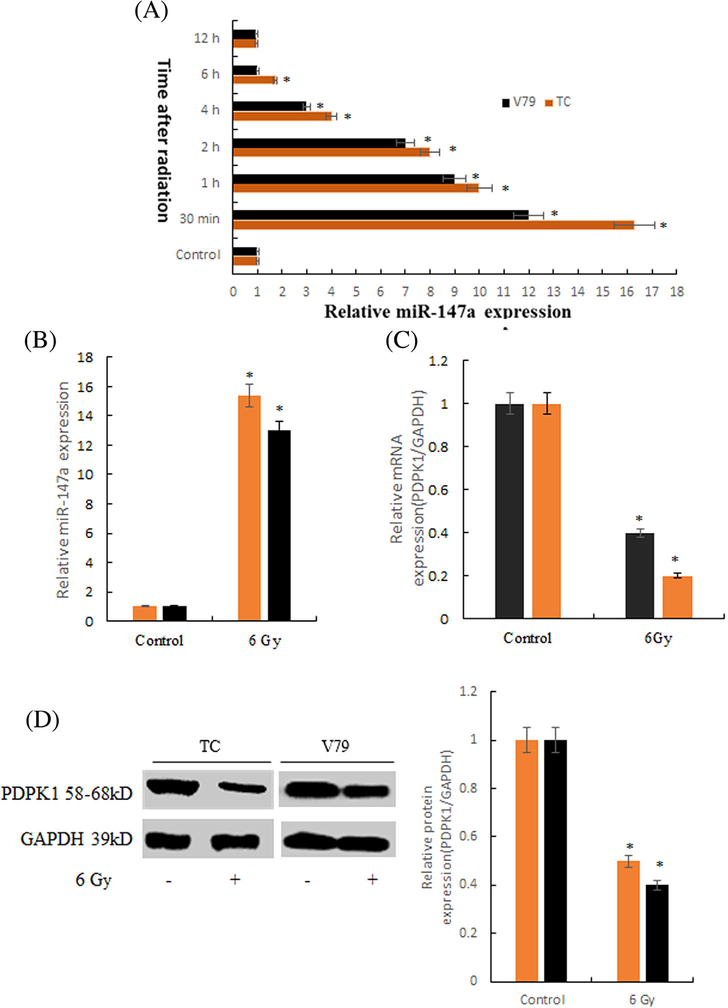
FIGURE 1.
The expressions of miR-147a and PDPK1 after irradiation in thymus cells. A, The optimal detection time of miR-147a was 30min after radiation (*P < 0.01 compared with Control). B, The expression level of miR-147a after radiation (*P < 0.01 compared with Control). C, The expression level of PDPK1 mRNA after radiation (*P < 0.01 compared with Control). D, The expression level of PDPK1 protein after radiation (*P < 0.01 compared with Control)

