Figure 2.
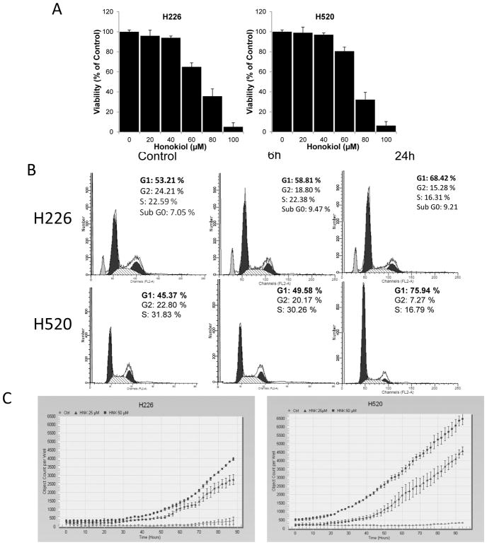
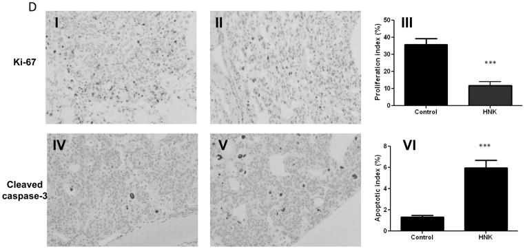
Effect of honokiol on cell proliferation, cell cycle progression and apoptosis in vitro and in vivo. A, Relative cell proliferation rate of H226 and H520 cells treated with honokiol at various concentrations. B,. Effect of honokiol on cell cycle arrest. C, Effect of honokiol on cleaved-caspase 3/7 staining. Shown is the caspase-3/7 fluorescence objects count from Incucyte. Data shown are the means ± SD (n = 3 per treatment group) D, Effect of honokiol on cell proliferation and apoptosis in NTCU-induced lung scc model. Lungs harvested from mice on the 26 weeks in NTCU study (n = 5 mice/group) were fixed, and the FFPE slides were stained using specific antibodies as detailed in the Materials and Methods section. I, II: Representative picture from immunohistochemistry for Ki-67 (I, control group; II, Honokiol group); III: proliferation index as determined by Ki-67; IV, V: Representative picture from immunohistochemistry for cleaved caspase-3 (III, control group; IV, Honokiol group). VI, apoptotic index as measured by cleaved caspase-3. ***, P < 0.001, control group versus honokiol group.


