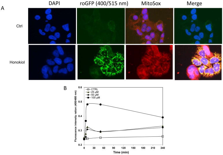
Figure 5.
Honokiol treatment shifts the mitochondria to a more oxidizing state. A, H520 cells with stable expression of pEGFP-mito-roGFP was stimulated with honokiol, Mito-EGFP- expressing H520 cells were loaded with MitoSOX Red (5 μM) for 20 min. roGFP states the production of ROS in mitochondria, which was also confirmed by MitoSOX red. Merged image shows co-localization of the mito-GFP fluorescence and MitoSOX Red (Mag. 40X). B, Honokiol promoted ROS production in NSCLC cells. Dual-excitation ratio measure (400/515nm and 480/515nm) with fluorescence plate reader was used to calculate ratiometric response from H520 cells after various concentrations of honokiol treatment at different time point.

