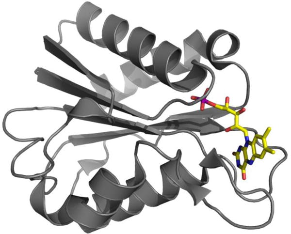
Figure 1.

Ribbon structure of FD depicting elements of secondary structure with ribbon segments and intervening stretches of backbone with cord. Amino acid side chains are omitted for simplicity however the flavin is shown in atomic detail with N atoms in blue, O atoms in red and the P atom in purple. H atoms are not shown and C is in yellow consistent with FMN’s intense yellow color. Figure generated using Pymol41.
