
Table 1.
Activity of Compounds 1–3 against DENV-2 Replication and DENV-2 NS5 RdRp.
| DENV-2a |
||||||
|---|---|---|---|---|---|---|
| RNA |
NS5 RdRp |
|||||
| Structure | CC50b(μM) | EC50c ± SD (μM) | SId | Cell-based EC50e ± SD (μM) | Enzyme-based EC50f ± SD (μM) | |
| 1 | 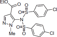 |
196 | 11.7 ± 0.2 | 16.7 | 8.1 ± 0.3 | 7.8 ± 0.3 |
| 2 | 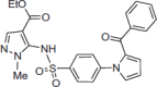 |
>200 | 7.6 ± 0.4 | >26.3 | 7.2 ± 0.4 | 5.3 ± 0.2 |
| 3 | 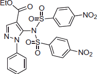 |
>200 | 5.7 ± 0.3 | >35.1 | 6.0 ± 0.3 | 4.9 ± 0.2 |
Data are mean values of two to three independent experiments each one in triplicate.
CC50: half maximal cytotoxicity concentration.
EC50 (DENV-2 RNA): half maximal effective concentration. Huh-7 cells were infected with DENV-2 and followed by RdRp inhibitors treatment for 3 days. The cell lysates were collected to analyse DENV RNA synthesis by qRT-PCR with specific primers targeting NS5.
SI: selectivity index calculated as CC50/EC50 ratio.
EC50 (cell-based DENV-2 NS5 RdRp): cell-based RdRp reporter assay. The Huh-7 cells were transiently expressed p(+)RLuc-(–)DV-UTRΔC-FLuc and DENV NS5 expression vector pcDNA-NS5-Myc.
EC50 (enzyme-based DENV-2 NS5 RdRp): enzyme-based RdRp activity assay. The (−) 3′UTR RNA was incubated with RdRp polymerase protein and CTP, GTP, UTP and BBT-ATP. The fluorescence signal was measured at excitation wavelength of 422 nm and emission wavelength of 566 nm, respectively.