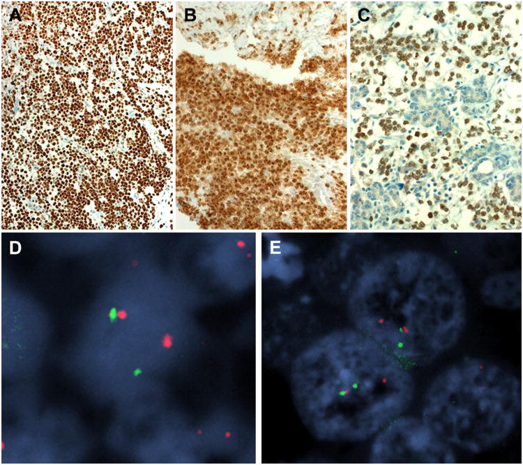
Figure 3.
Immunohistochemistry showed consistent expression of p63 (A; Case 3) and NUT protein (B; Case 2). The NUT immunostain highlighted the neoplastic cells amid native salivary tissue (C; Case 3). FISH analysis using the NUT probe (D) and BRD4 probe (E) showed break-apart signals indicating a NUT/BRD4 translocation (image from Case 1).

