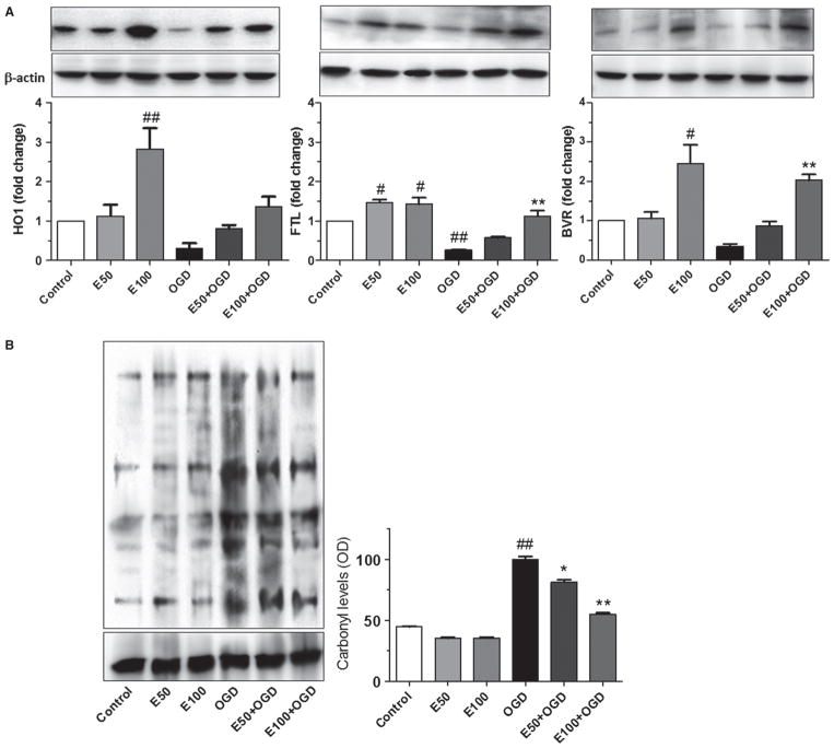
Fig. 3.
EC protection occurs through upregulated expression of Nrf2-responsive proteins and reduced protein oxidation. (A) Western blot shows dose-dependent increase in HO1, FTL, and BVR in WT neurons. (B) OxyBlot assay and quantification shows reduced carbonyl levels in neurons treated with 50 or 100 μM EC relative to untreated cultures. *P < 0.05; **P < 0.01; #P < 0.05; ##P < 0.01; #compared with control; *compared with OGD.

